Standard scanning electron microscopes
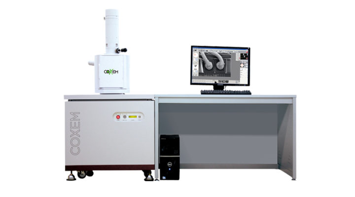
CX-200 Series
2. Full automatic functions (X, Y, R, T, Z axes) or partly manual
3. The large room can accommodate up to 160mm (W) * 55mm (H)
4. Magnification of x 300,000 (20nm resolution at 30kV)
5. Quick replacement of a sample in less than two minutes
High resolution standard SEM microscopes
The CX-200 series is a standard scanning electron microscope (SEM), suitable for both experienced and experienced users, for many types of research and quality control requirements.
The CX-200s offer high resolution imaging capabilities at a very attractive price. After evaluating the CX-200s, your search for the most cost-effective solution will be an easy decision.
Applications of Scanning Electron Microscopy
The applications include both the fault process, dimensional analysis, the characterization process, reverse engineering and particle identification ... this is why the fields of application are multiple and very broad:
- Life sciences
- Microbiology
- Food / Environmental Sciences
- Plants and animals
- Medicine / Pharmaceutical
- Human body
- Microbiology
- Materials science
- Chemistry
- Automobile
- Construction
- Smartphones
- Energy
- Semiconductors and electronics
- Metals
- Chemistry
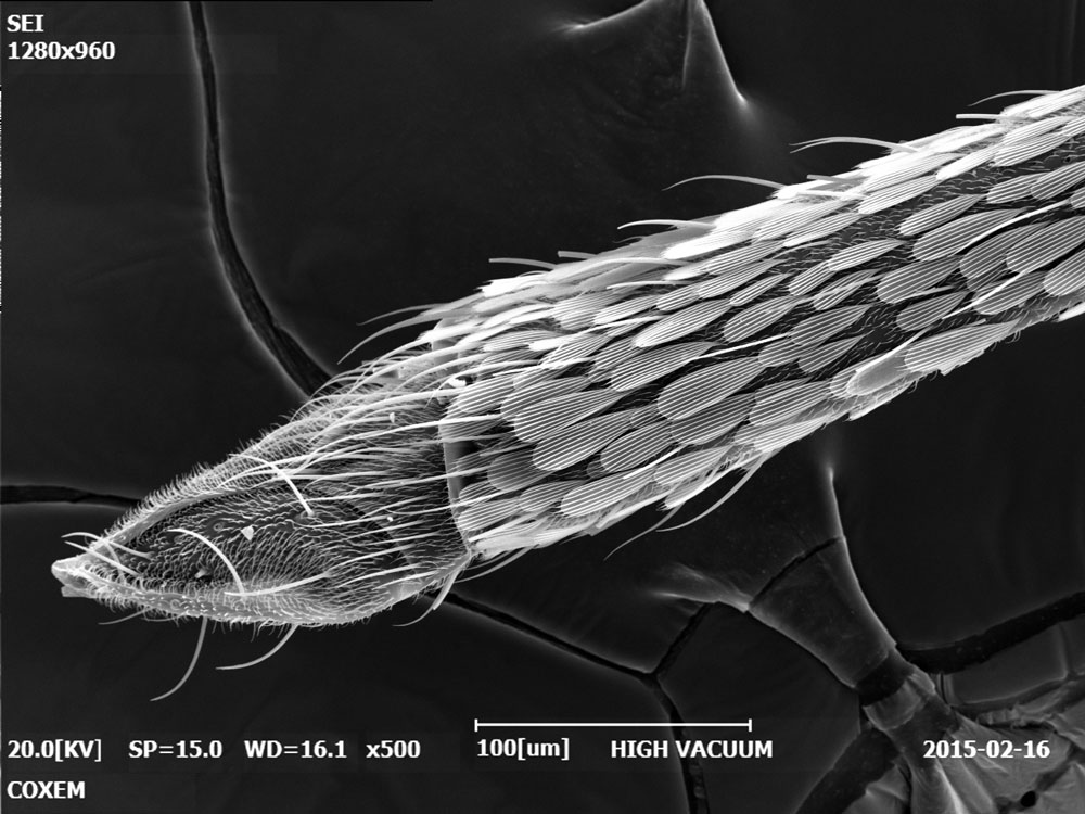
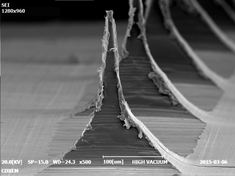
Mosquito stinger
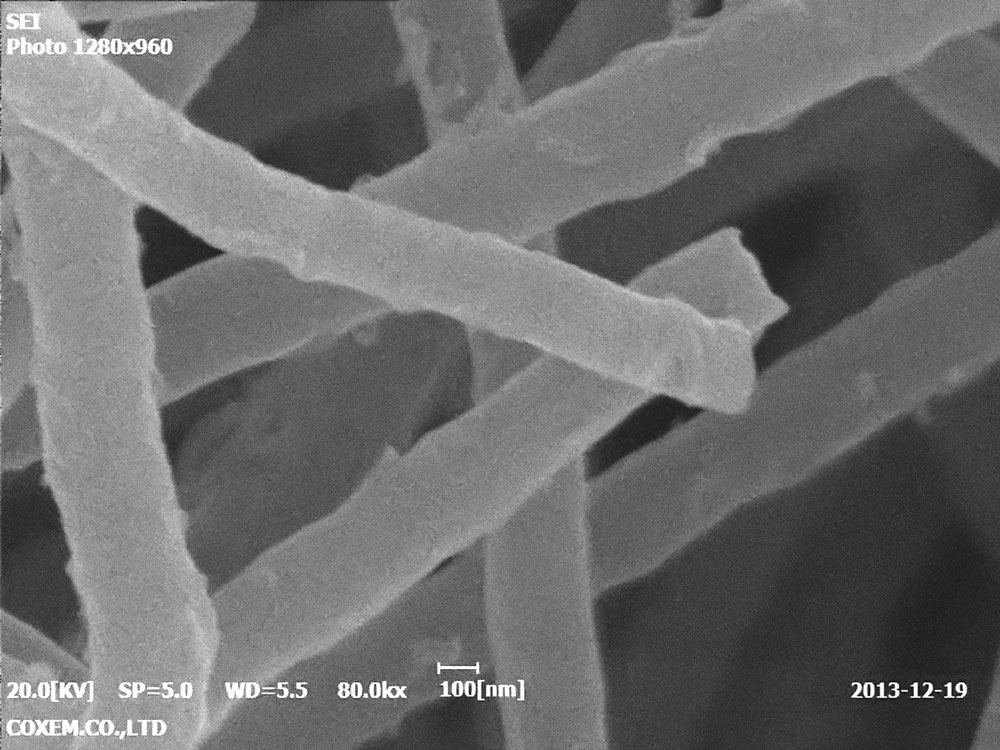
Nanotubes 80000x
Silicon wafer 2
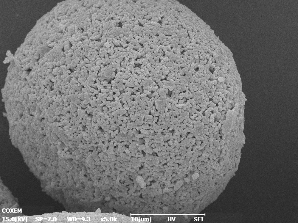
X2-5
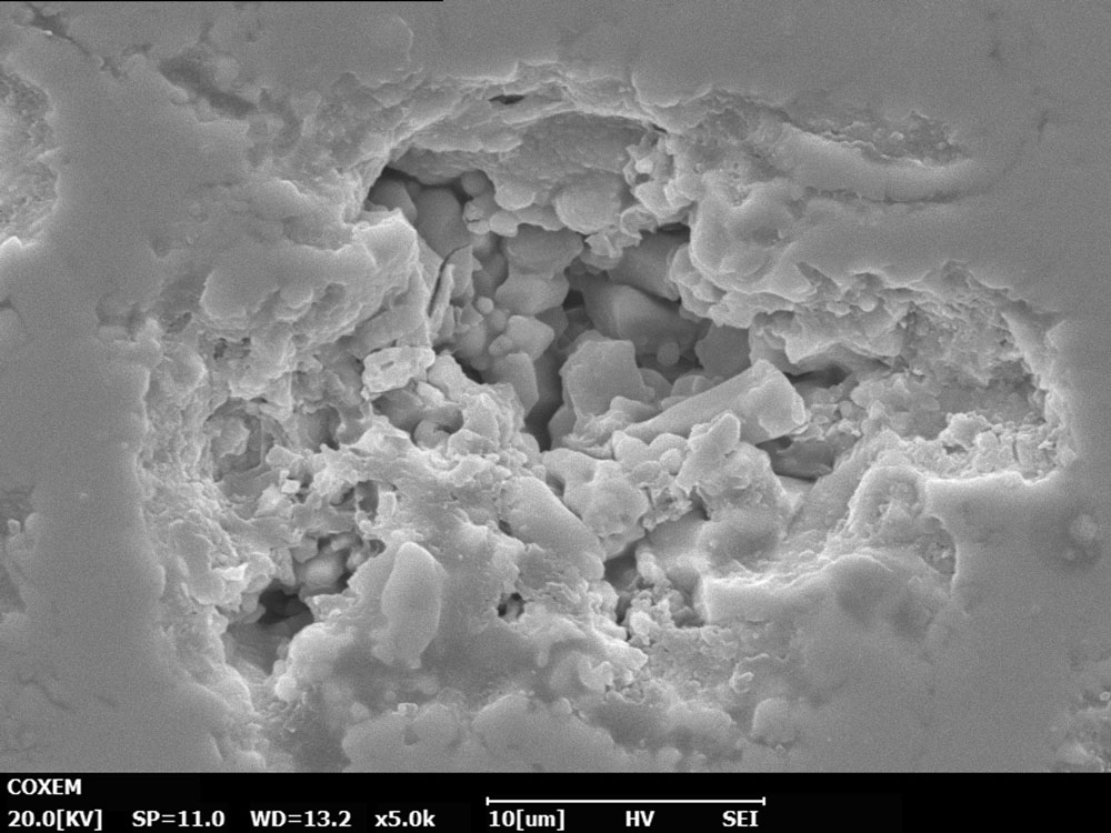
3-4
Cosmetic patch
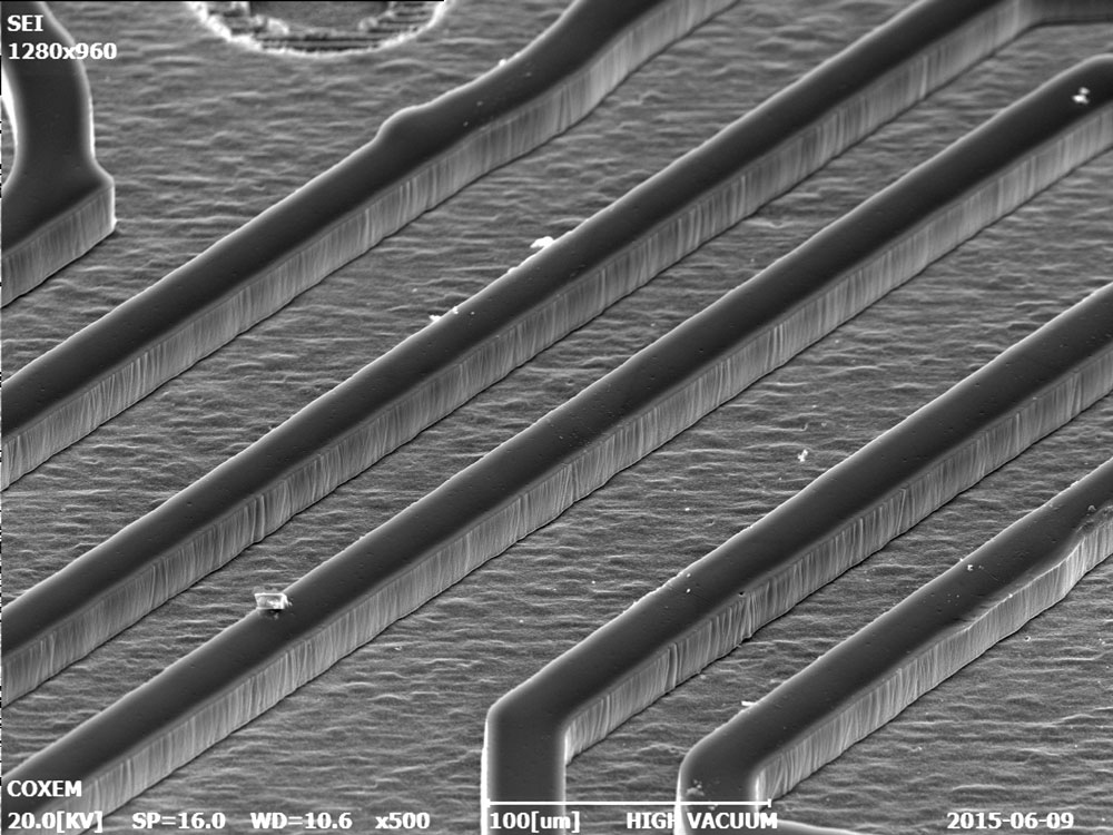
Etch-500x
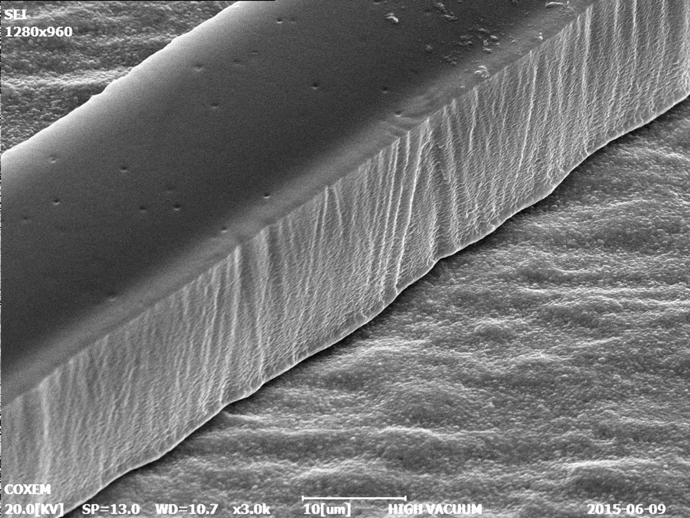
Etch-3000x
More info on applications
- High resolution
- SE + ESB mode
- Vacuum mode
- Acceleration voltage
- Angle of inclination
- Coolstage application
- Panorama Shot application
- Application of 3D topography software
- Application of transmission scanning electron microscopy (STEM)
- Application of EDS
- Cartographic analysis
- Large-scale mapping
- Analysis of gunshot residues (GSR)
- Multilayer analysis
- Mineral cartographic analysis
Scanning Electron Microscopy
high resolution at low cost
The CX-200s offer a unique “Panorama” mode which allows the SEM to collect high resolution images of large samples by controlling the motorized stage at high magnification and to automatically collect large images from several to thousands of images in mosaic. This feature is very useful for applications such as in-depth examination or metrology of semiconductors and electronic assemblies, as well as for the production of images that can be printed in poster or high definition wall format.
Optional analytical detectors:
Elementary analysis EDS and WDS - EBSD - Cathodoluminescence (CL) - SE / BSE
Optional accessories:
Sample holder - Cooling step - Plasma cleaning - Anti-vibration platform
Imaging capabilities
- Resolution: 3 nm
- Magnification: 300,000x
- Energy beam: 1 - 30 kV
- Sample positioning: 5 axes (XYZRT)
- Extensibility: 10 ports
ADVANTAGES
- Full automatic functions (X, Y, R, T, Z axes) or partly manual
- The large room can accommodate up to 160 mm (W) * 55 mm (H)
- Magnification of x 300,000 (20nm resolution at 30kV)
- Quick replacement of a sample in less than two minutes
- Working distance
- High resolution
- SE + ESB mode
- Vacuum mode
- Acceleration voltage
- Angle of inclination
- Coolstage application
- Panorama Shot application
- Application of 3D topography software
- Application of transmission scanning electron microscopy (STEM)
- Application of EDS
- Cartographic analysis
- Large-scale mapping
- Analysis of gunshot residues (GSR)
- Multilayer analysis
- Mineral cartographic analysis
SE (electronic secondary) and BSE (electronic backscatter) imagery
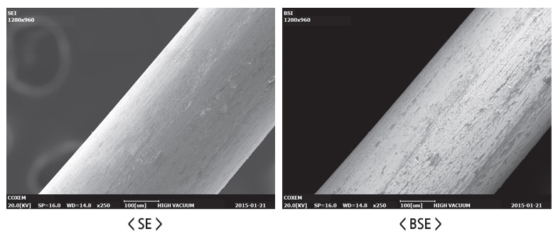
Screenshot with surface and topographic details (SE) or atomic weight contrast (BSE)

Combine the two imaging detector signals to obtain a composite image representing both the change in topographic and atomic weight
Functional and user-friendly functions
NANOSTATION SOFTWARE
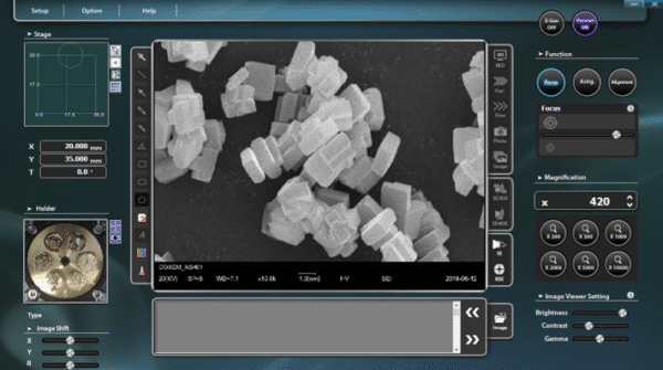
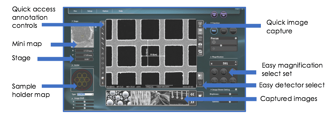
NanoStation software provides the novice user with a simple, clean, clutter-free interface, while the more advanced user can easily access features that allow more control over microscope functions. With the intuitive software, users can view and save images quickly and easily with great detail.
The user can select a standard sample holder layout at the bottom left or use the digital “NavCam” to capture an image of the sample or media to be used for macro navigation. In the upper left corner of the interface, the mini-map is an image collected at the lowest magnification desired for the field of vision or the area of interest. When the user navigates to another location on the sample and at higher magnifications, the location of the current view is indicated on the mini map (yellow rectangle).
All "slide" controls can be easily adjusted using a variety of techniques: dragging the indicator, using the mouse wheel, a +/- button, the keyboard, and some "auto" adjustment functions.
In addition to the keyboard shortcuts preferred by advanced users for certain functions such as focusing, the software provides easy access to all microscope and imaging settings by mouse click or mouse wheel. All the function control groups are intelligently organized on the software interface. The less common parameters, generally defined according to user preferences, are invisible in the secondary windows.
NAVIGATION
The EM-30 table-top SEMs include a variety of sample navigation tools to assist new and experienced users alike. A digital "NavCam" can be used to capture an image and / or a sample holder, which is then placed in the sample card area of the software GUI. NavCam is standard on the EM-30 LE and optional on the EM-30 Plus. Alternatively, a variety of multi-sample presentations can be selected from a drop-down menu which is conveniently used to move from one sample to another.
It is very easy to navigate through different samples or different areas of a sample using a variety of commands to suit the preferences of each user.
- Click with the mouse on any sample of the NavCam image or on the card of the sample holder to access this sample.
- Click with the mouse on an entity observed in the mini-map to center the radius on this entity.
- Click anywhere in the largest scanned image to move this point to the center of the beam
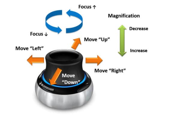
- Use the supplied joystick (shown on the right) to "drive" the scene in any direction, zoom in / out or adjust the focus.
- The arrow keys on the keyboard are used to control the scene or the image to the left / right and up / down.
- The X-Y coordinates of known entities can also be entered to direct the beam to these coordinates
- At very high magnification, the software's Beam Shift sliders are used to make micro and nano movements
SAMPLE POSITIONING
The EM-30 series desktop scanning electron microscopes come with a standard 3-axis motorized stage for X / Y and Tilt. The working distance or "Z" is very easy to define before closing the chamber and avoids accidental collisions in the absence of an external Z axis.
The Tilt function is extremely useful for examining the topography of flat samples. The tilt function is complex, so the X axis is adjusted during tilt to keep the region of interest of the sample in the field of view. The compucentric tilt function offers more versatility and time savings thanks to its easier use, especially for inexperienced users.
PRESENTATION EXAMPLE
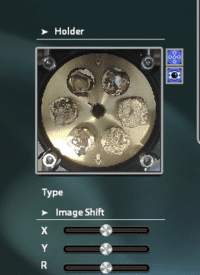
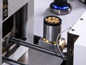
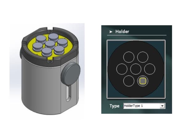
Sample mounting is easily accomplished using various sample holders provided, which allow the use of standard pins of various sizes. The standard multi-pin holder (shown on the right) allows easy mounting of up to 7 bits of 6 mm in diameter, 3 bits of 12.7 mm or 1 bit of 25 to 32 mm in diameter. The multi-support can be easily adjusted to the height in Z using the side knurled screw in order to receive large samples or other supports such as small vice clamps or pre- tilt of various suppliers - LINK.
The type of sample holder is selected in the software which presents a sample card in the lower left corner of the interface, which allows to easily switch to different samples without losing track of the sample being created. 'picture. Alternatively, the digital "NavCam" can be used to capture an image of the sample and / or the medium to be inserted there.
Adapters are also provided to allow the attachment of one of your existing supports with an M4 thread. If you prefer brackets other than pins such as Hitachi M4 style fasteners or Jeol style cylinder fasteners, our staff have extensive experience in adapting many types of mounting systems for SEM systems. So please call on us.
PANORAMA MODE - Large format image capture
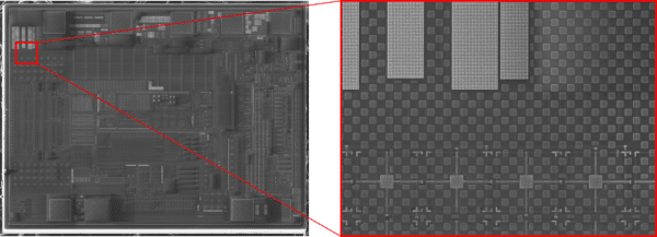
NanoStation 4.0 software provides the ability to capture high resolution images of a larger area by automatically moving the scene to capture a few individual images in a checkerboard pattern, which are then automatically assembled to form a large image the size of a giga-pixel. . You can also save individual images for batch download in image processing software such as MIPAR or ImageJ to quantify particle size, cells, fibers, body shape and more.
The process of capturing large images is quite simple and requires only one menu for configuration. The progress of the image captures is displayed in the mini-map area at the top left.
This larger giga-pixel image can be used for a variety of purposes, such as printing a high-resolution wall-size image, for example. For semiconductor circuits, a single image allows the analyst to pan and zoom in on a single image for an inspection role such as fault analysis.
BACKSCATTER ELECTRON DETECTOR (BSED)
The EM-30 Plus series scanning electron microscopes (SEM) are delivered with a 4-quadrant backscatter electron detector that can be used in COMPOSITION or TOPO mode. Composition mode is standard for atomic contrast images, useful for both metrology measurements and EDS. Topo mode is a synthetic image created by alternating the quadrants of the detector. This can be useful for topographic-looking images on flat samples with sample tilt.
The BSE detector is also retractable without disconnection - a unique feature not found in other table or office SEMs. The BSE sensor can be swiveled from the SEM pole piece to a parking position. This allows unobstructed access to shorter working distances using the SE sensor and for other purposes.
IMAGE METROLOGY TOOLS
Metrology or image measurements are extremely flexible and precise with the EM-30 series from SEM. Extended calibration is performed at the factory at multiple magnifications, working distances, beam energies and more, resulting in measurement errors of less than 1%. Detailed calibration reports accompany the documentation for each system, making the EM-30 Plus a valuable tool that can be easily certified and accepted in your workflow.
The software provides quick access to all common measurement tools such as:
- Length
- Diameter
- Area (rectangle, circle, polygon)
- Angles
- Annotation
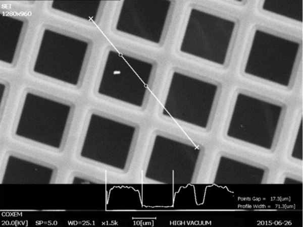
In addition, a line scan function (shown at right) can be used for very detailed and precise measurements using the variation in brightness of each pixel to accurately determine the true location of edges and features.
Models
Specifications Standard SEM
2 models of standard scanning electron microscopes are available:

Contact us for more information on this product
Would you like an estimation ?
Additional information?
We will reply to you within 24 hours


